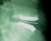
Found those threads, and the before x-ray, I can't really tell anything was wrong; but it shows the progression. http://www.ispine.org/forum/ispine/1...dr-stenum.html
Here is the other one:
http://www.ispine.org/forum/surgical...um-kc0iet.html
On another question. Have you/did you follow all the post surgery orders? Not sure the exact ones for cervical; but for lumbar it was no bending, twisting, lifting, etc. Did you fall or anything? Just curious, as I am terrified of my discs moving out of place. I am always relieved to see my new images, to know everything is where it is supposed to be. Oh, do you have any of your earlier images? Do they look like this? Just curious.
Oh, just read this in the old post, what Mark said (mmglobal) regarding the discs being gone, which takes the pain away; but is still not a 'success'. Here, I am copying and posting a portion of that post, as he explained it better than I can. (underlining mine)
Quote:
Originally Posted by mmglobal

"First, notice the angulation of the Charite’ plates on the first picture. The implant is not centered well. This is causing the upper plate to ‘fall off’, lower on the left side of the image. This demonstrates one of the problems with mobile core devices. When this occurs, the core is pushed to the extreme right and stays there. That increases the angulation and increases the forces that push the core more off center in the wrong direction. This is why the Activ-L eliminated the lateral movement of the core. Every mobile core device I’ve seen will do this. Properly implanted it’s much less of an issue. I’ve had 2 clients with M6 cervical discs explanted, one for problems much like I just described, another one for serious complications that may have been exacerbated for these reasons. (Yes, they were both Stenum patients. I know of a third, but I was not involved in the case. I did get to examine the explanted disc though.)
That brings us to the second picture. Notice how far the back of the upper plate is from the back of the vertebral body. Notice how the teeth of the plate are literally on top of the anterior margin of the vertebral body. There is the appearance of more vertebra because of an anterior osteophytes. This kind of alignment increases the risk of migration by many orders of magnitude. I see these types of films presented at the conferences as if they are a device issue, but this is not a device issue. The picture of the configuration before migration is one of a disaster waiting to happen. The surgeon should know that and be focused on proper placement. The doctors at Stenum say that there are anatomical reasons that may make it impossible to get the disc further back. That is absolute BS. I have NEVER seen this type of failure from any of the other surgeons I work with because they take care to get it right. Accepting sloppy work because you are lazy, hurried or just not careful may not cause problems most of the time. However, when the stakes are soooooo very high, accepting sloppy work may doom patients that would have otherwise been successful, to lives of pain, meds, revision surgeries and more.
After I went to Stenum with MrBee, I made excuses for them, saying that they are probably doing the surgery the way they were taught to do it years ago. The reply from one of my favorite surgeons was, “If you are a bricklayer or a librarian, that may be OK. But if you are an astronaut, an airline pilot, race care driver or a surgeon, you have to be learning all the time. That is not an excuse.”
Here is a picture that I extracted from the original Stenum-and-back website. This picture stayed up there for many years until the patient community got wise to what it really showed.

I want everyone to keep in mind that this is the image of a successful surgery. The author of the website may even be in better shape than me. They point to images like this as if it’s evidence that it’s OK to do surgery this way. However, you do NOT want any ADR implanted this way. If the patient’s disc was his pain generator and they took it out, he experiences success. If he gets away with the horrible placement, that is dumb luck, not appropriate surgical technique. At the conferences they discuss the sequelae of configurations like this: increased risk of complications like migration and subsidence. In addition, there is the expectation of accelerated wear and degeneration of posterior elements, possibly adjacent levels (due to inappropriate kinematics), AND of the prosthesis itself. It’s like driving with your tires out of alignment.
Does this mean it WILL happen? Absolutely not! All it does is increase your risk. Poor surgery does not guarantee failure just as perfect surgery does not guarantee success. If anyone wants to have poor surgery because it’s OK most of the time, I would suggest that they don’t fully understand the issues." |
__________________
34 years old-
1/06- In wreck with 18 wheeler
Numerous MRI's, PT, chiropractic, accupuncture, TENS therapy, massage therapy, facet injections, epidural injections, Nerve study, Discogram, confirms pain in L4/5, IDET, decompression, Bi-lateral neurotomy L3/4/5, denied by insurance twice, in Active L clinical trial, had surgery March 17, 2009 in Miami, FL- received Active L disc
Had Baby #3 after ADR!
Last edited by Kathy; 05-17-2009 at 03:29 PM.
|